
Olga Klachkovich, PA-C &
Venu M. Nemani, MD, PhD
Virginia Mason Medical Center
Osteoporosis is a common condition that is characterized by a low bone mineral density leading to increased risk for fractures (broken bones). Your healthcare provider can use several diagnostic tests and tools to screen for osteoporosis. The three tests reviewed below complement a complete history and physical examination, which includes blood work to assess your clinical risk factors.
DXA
Dual energy x-ray absorptiometry (DXA) provides a measure of bone mineral density by passing both high-energy and low-energy x-ray beams through various parts of your skeleton, usually your hip (proximal femur), spine, and sometimes your forearm/wrist (distal radius). This is the
most common noninvasive screening test for osteoporosis that is quick, painless, and with low exposure to radiation. DXA scans should be repeated once every 2 years or sooner if there has been a clinical change that might adversely affect bone density (e.g., starting chronic high-dose steroids).
DXA reports bone density as a “T-score,” which compares your bone density to that of a healthy young adult. A T-score above -1 indicates normal bone, a T-score between -1 and -2.5 indicates osteopenia, and a T-score less than -2.5 indicates osteoporosis. The lower your score, the weaker your bone. It is important to note that if you have suffered a fracture due to a fall from a standing height or a similar low-energy injury, then by definition you have osteoporosis regardless of your T-score on a DXA scan.
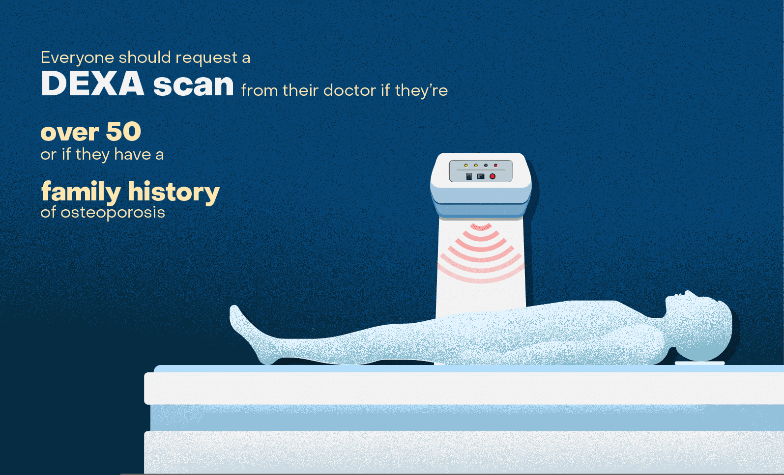
DXA scans can be done at most radiology imaging centers and can be ordered by your primary care provider or specialty providers such as a physiatrist, endocrinologist, or your spine surgeon. Screening with a DXA scan should be considered in:
- All post-menopausal women 65 years or older
- Men greater than 70 years old
- Men and women 50 years or older with known risk factors for osteoporosis, such as:
- Smoking/nicotine use
- Vitamin D deficiency
- Prolonged use of steroids
- Family history of osteoporosis or a parent with hip fracture history
- Malabsorption
- Chronic illnesses
Additionally, we strongly recommend screening with DXA in all patients who are being considered for spinal fusion surgery (especially multi-level spinal fusion) who have risk factors or fracture history, so that low bone density can be optimized pre- and post-operatively if identified. This can significantly lower the risk of osteoporosis-related complications, surgical failures, and the potential need for revision (redo) surgery. Finally, a lateral spine X-ray can be obtained at the time of DXA (called vertebral fracture assessment or VFA) which is used to identify occult spinal fractures. This is especially important in patients about to undergo spine fusion.
FRAX
There are several online tools and calculators that allow you or any clinician to input your clinical information and estimate your personal risk for osteoporosis or risk of fractures. One of the best tools available to assess fracture risk is called the FRAX calculator. This tool gathers responses to 12 questions related to your clinical risk factors including age, gender, weight, smoking history, alcohol use, and other factors that can affect your bone health. It can be used by anyone at home, with or without DXA data. It provides you with a 10-year probability of a hip fracture and a 10-year probability of a major osteoporotic fracture (fracture in your spine, hip, shoulder, or forearm). These fractures can be devastating and significantly impair one’s quality of life.
FRAX results are expressed in percentages. A 10% risk of a hip fracture, for instance, would mean that 10 out of 100 people with the same risk profile as you will develop a fracture in the next 10 years. If your FRAX results give you a 10-year probability of a hip fracture > 3%, or major osteoporosis-related fracture > 20%, then you should talk to your healthcare provider about starting prescription medications to treat your osteoporosis. Calcium and Vitamin D are not enough to lower your risk of fracture in these cases!
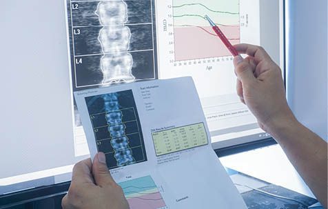
Figure 1 is an example of the important additional information the FRAX tool provides. In this case, the person’s DXA results (T=-2.1) put them in the category of osteopenia (no need for prescription treatment), but FRAX results considered their clinical risk factors and put them at high risk for fracture (prescription treatment needed).
Opportunistic CT Scans
Another way your provider or spine surgeon may diagnose low bone density is by using information from a CT scan you’ve already had, called an “opportunistic CT scan.” A CT evaluation of your lumbar or thoracic region may be requested by your spine surgeon as part of the pre-operative planning process for spine surgery. From that CT scan, your surgeon can additionally measure what are called “Hounsfield Units” (HU). This evaluation provides a measure of local bone density that can be specific to the spinal levels where you may be having surgery, rather than a regional evaluation of bone from DXA. Generally, an HU below 110 correlates with the presence of osteoporosis.
Your provider can measure HU numbers not only from a dedicated spinal CT study but also from a CT of the chest, abdomen, or pelvis that may have been ordered for other clinical reasons. Although HU measurements alone cannot be used to diagnose osteoporosis, when combined with other clinical and diagnostic data, they can support the approval of osteoporosis treatment if your surgeon or primary care provider deems it necessary for optimization of your bone health.
Summary
Poor bone health is a common medical problem without obvious symptoms to prompt you to seek workup and treatment until late in the disease process when fractures occur. The three tools described in this article are noninvasive and can provide a wealth of knowledge for you both now as a baseline and in the future as your bone health changes. Adults, particularly those with risk factors, should be proactive in asking their healthcare providers to evaluate their risk of osteoporosis by these easy and effective clinical tests so that education and the appropriate treatments can be initiated expediently.