Nathan J. Lee, MD and Ronald A. Lehman Jr., MD

The adult spinal deformity (ASD) population is incredibly diverse, making it critical that surgical treatment is individualized for each patient. Several factors should be considered when deciding on the optimal surgery. A thorough evaluation of the medical history, symptoms, physical examination, and imaging studies are essential to determine the most appropriate treatment plan. Furthermore, it is absolutely essential to involve the patient in the decision-making process. Realistic discussions about potential benefits, risks, and limitations of surgical interventions should take place preoperatively to set appropriate goals and expectations. Several of these conversations may be necessary over time, given the complexity of the spinal condition, the surgical treatment, the recovery, and life after ASD surgery.
Types of Surgery
Although surgical treatments for ASD may differ between younger and older patients, these differences depend less on the patient’s actual age and more on the variations in bone quality, overall health, radiographic spinal phenotype, symptomatology, and the patient’s lifestyle. Depending on these factors, an ASD patient may require either decompression alone, decompression and fusion, and/or a realignment procedure.
- Decompression (creating more room for neural structures by removing compressive bone/tissue) may be used alone for patients with a primary complaint of stenosis (narrowing around neural structures) who have a stable and well-balanced spine.
- In patients with destabilizing pathology (such as slipping of the vertebrae = spondylolisthesis), a fusion procedure (taking away motion of spinal segments) should be included. Decompression without stabilization in patients with instability may further destabilize the spine and lead to deformity progression.
- Realignment procedures are appropriate when the primary aim is to correct the deformity, improve global and regional alignment, and prevent progression. However, realignment surgery is more extensive in nature as it includes decompression, fusion, and osteotomies (a surgical technique to loosen parts of the spine to allow for realignment in an otherwise stiff area).
The Importance of Imaging for Realignment
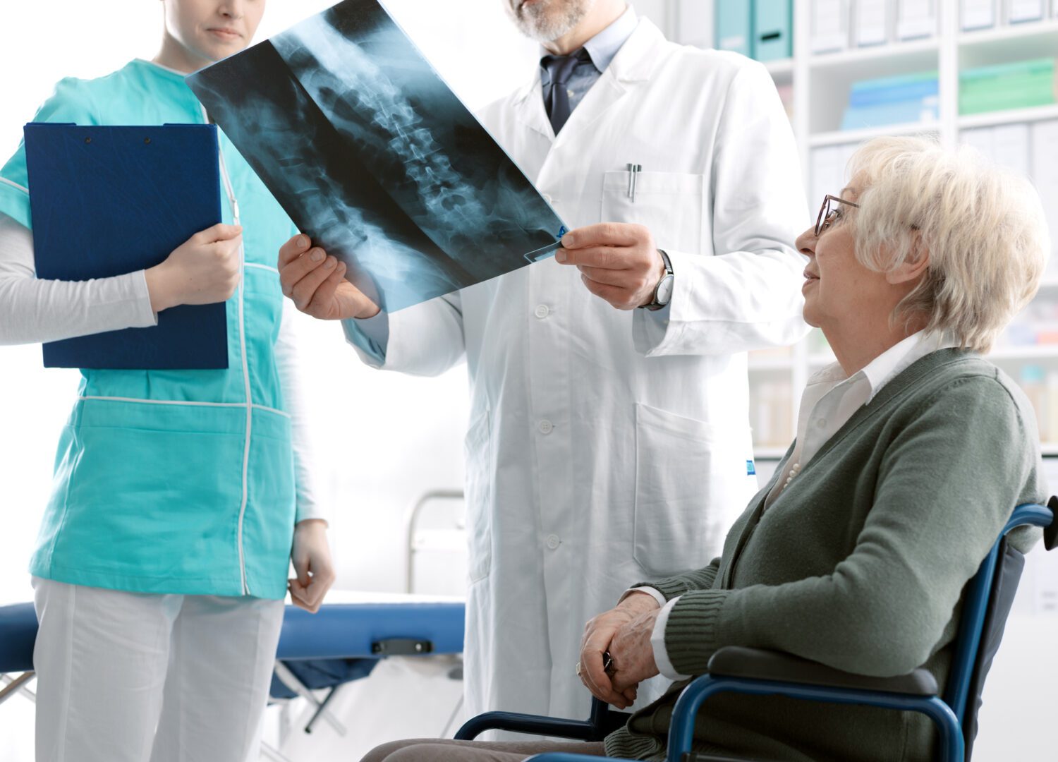
In addition to the well-known evaluation using magnetic resonance imaging (MRI), multiple radiographic factors are important to consider in determining the optimal surgical treatment for younger and older patients alike. First, full-length standing x-rays are used to assess alignment in the sagittal (side) and coronal (front-back) planes. Patients may appear globally well-aligned on standing x-rays, but special attention should be made to any compensatory changes made in the pelvis and legs, which may mask the true alignment. There are also natural curves in the spine seen from the side view that balance each other for standing upright: lordosis in the neck, kyphosis in the thoracic region, and lordosis in the low back.
These natural curves can change over time and contribute to a spinal deformity condition. The magnitude of mismatch between what the natural curvature in the low back should be and what it has become over time is particularly important in patients who develop a forward-flexed posture. Surgical correction of this mismatch improves posture and is associated with improved patient-reported outcomes.
Flexibility x-rays include laying or bending films to assess the rigidity of the thoracic and lumbar curves. CT scans can further characterize the rigidity of curves and illustrate abnormal bone growth, facet joint arthritis, and lumbar degeneration. In some cases, rigid curves may require extensive bone work to loosen up the spine for correction, including:
- Pedicle subtraction osteotomies (PSO)
- Vertebral body resection (VCR)
These types of three column osteotomies (3CO) can achieve greater correction of spinal deformities, but are associated with greater blood loss, prolonged operative time, risk for neurologic deficit, and pseudarthrosis (failure to fuse).
Level Selection for Realignment
The number of spinal levels included in ASD surgery must be carefully considered. The first (or top) level is known as the upper instrumented vertebra (UIV) and the last (or bottom) level is known as the lower instrumented vertebra (LIV). The appropriate determination of the UIV and LIV is critical to avoid potential postoperative complications such as new issues developing at the levels above and/or below the surgical levels or a failure to fuse.
The Lenke classification is often utilized for optimizing level selection. Ultimately, level selection should be based on coronal and sagittal alignment, relative magnitude and rigidity of curves, the quality of the muscles that surround the spine, shoulder imbalance, degree of lumbar disc degeneration, and how even the hips/pelvis are. Careful consideration should be made to avoid ending the fusion construct at locations known to be less stable or anatomically problematic.
Surgical Approach to Realignment
An open, posterior-based (long incision down the middle of the back) deformity correction is considered the gold standard surgical approach for ASD. The main advantage is the ability to correct large deformities with a single approach. All concomitant procedures including interbody fusions and osteotomies can be made within the same incision. However, this approach is associated with relatively high complication rates.
To mitigate these risks, minimally invasive spine surgery (MIS) has been introduced with the primary goal of preserving native soft tissue anatomy, which is disrupted during an open approach. Other benefits of MIS include less blood loss and faster recovery. The indications for the MIS techniques continue to evolve. MIS may be best suited for patients with flexible curves of the thoracolumbar/lumbar spine. MIS is relatively contraindicated in patients with very rigid and large curves or those with significant fixed sagittal imbalance.
Innovative Surgical Technology
Significant technological advances have been made in spine surgery over the last decade, including robot-assisted spine surgery, navigation, and machine learning. Recently, robot-assisted spine surgery has been integrated with navigation technology, which has been shown to reduce radiation exposure and improve instrumentation accuracy. An important advantage of robot-assisted technology is the improved ability to preoperatively plan the surgery, allowing for individualized plans based on each patient’s unique needs. Clinical research has demonstrated promising outcomes with the use of this technology. Furthermore, the use of machine learning to further personalize care has continued to expand over the last several years.
Conclusion
Ultimately, the optimal surgical treatment for ASD will rely on a transparent preoperative discussion with the patient to foster shared surgical decision-making. Surgeons should account for patient factors, including lifestyle choices, comorbidities, and their radiographic phenotype, as well as set realistic short- and long-term goals of surgery. Through this discussion, surgeons should be able to determine which tools to use in their armamentarium to provide individualized care. No two ASD patients are alike!